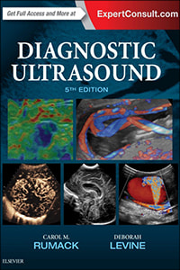
Now fully updated with more than 2,000 new images, 200 new videos, and new content throughout, Diagnostic Ultrasound, 5th edition, by Drs. Carol M. Rumack and Deborah Levine, remains the most comprehensive and authoritative ultrasound resource available.
Contents
Overview
Spanning a wide range of medical specialties and practice settings, it provides complete, detailed information on the latest techniques for ultrasound imaging of the whole body; image-guided procedures; fetal, obstetric, and pediatric imaging; and much more.
Up-to-date guidance from experts in the field keep you abreast of expanding applications of this versatile imaging modality and help you understand the “how” and “why” of ultrasound use and interpretation.
Features:
- Covers all aspects of diagnostic ultrasound with sections for Physics; Abdominal, Pelvic, Small Parts, Vascular, Obstetric, and Pediatric Sonography.
- Uses a straightforward writing style and extensive image panels with correlative findings.
- Features 5,000 images – more than 2,000 brand-new – including new 2D and 3D imaging as well as the use of contrast agents and elastography.
- Includes a new virtual chapter on artifacts with individually labelled images from throughout the book, displaying artifacts with descriptive legends by category and how they can be used in diagnosis or corrected for better quality imaging.
- Features more images and new uses for contrast agents in the liver, breast, and in pediatric applications. Includes current information on imaging more diagnostic dilemmas, such as Zika virus in the fetus and newborn.
- Includes 400 video clips showing real-time scanning of anatomy and pathology.
Explore more books:
Book Details – Diagnostic Ultrasound 5th Edition
Book Details

- Title: Diagnostic Ultrasound 5th Edition
- Author: Carol M Rumack Deborah Levine
- Publisher: Elsevier
- Publication: 2018
- Edition: 5th Ed
- Language: English
- Book Format: PDF
- Book Series:
Table of contents
- Cover image
- Title Page
- Table of Contents
- Copyright
- About the Editors
- Contributors
- Dedication
- Preface
- Acknowledgments
- Videos
- Part I Physics
- Chapter 1 Physics of Ultrasound
- Chapter 2 Biologic Effects and Safety
- Chapter 3 Contrast Agents for Ultrasound
- Part II Abdominal and Pelvic Sonography
- Chapter 4 The Liver
- Chapter 5 The Spleen
- Chapter 6 The Biliary Tree and Gallbladder
- Chapter 7 The Pancreas
- Chapter 8 The Gastrointestinal Tract
- Chapter 9 The Kidney and Urinary Tract
- Chapter 10 The Prostate and Transrectal Ultrasound
- Chapter 11 The Adrenal Glands
- Chapter 12 The Retroperitoneum
- Chapter 13 Dynamic Ultrasound of Hernias of the Groin and Anterior Abdominal Wall
- Chapter 14 The Peritoneum
- Chapter 15 The Uterus
- Chapter 16 The Adnexa
- Chapter 17 Ultrasound-Guided Biopsy of Chest, Abdomen, and Pelvis
- Chapter 18 Organ Transplantation
- Part III Small Parts, Carotid Artery, and Peripheral Vessel Sonography
- Chapter 19 The Thyroid Gland
- Chapter 20 The Parathyroid Glands
- Chapter 21 The Breast
- Chapter 22 The Scrotum
- Chapter 23 Overview of Musculoskeletal Ultrasound Techniques and Applications
- Chapter 24 The Shoulder
- Chapter 25 Musculoskeletal Interventions
- Chapter 26 The Extracranial Cerebral Vessels
- Chapter 27 Peripheral Vessels
- Part IV Obstetric and Fetal Sonography
- Chapter 28 Overview of Obstetric Imaging
- Chapter 29 Bioeffects and Safety of Ultrasound in Obstetrics
- Chapter 30 The First Trimester
- Chapter 31 Chromosomal Abnormalities
- Chapter 32 Multifetal Pregnancy
- Chapter 33 The Fetal Face and Neck
- Chapter 34 The Fetal Brain
- Chapter 35 The Fetal Spine
- Chapter 36 The Fetal Chest
- Chapter 37 The Fetal Heart
- Chapter 38 The Fetal Gastrointestinal Tract and Abdominal Wall
- Chapter 39 The Fetal Urogenital Tract
- Chapter 40 The Fetal Musculoskeletal System
- Chapter 41 Fetal Hydrops
- Chapter 42 Fetal Measurements
- Chapter 43 Sonographic Evaluation of the Placenta
- Chapter 44 Cervical Ultrasound and Preterm Birth
- Part V Pediatric Sonography
- Chapter 45 Neonatal and Infant Brain Imaging
- Chapter 46 Duplex Sonography of the Neonatal and Infant Brain
- Chapter 47 Doppler Sonography of the Brain in Children
- Chapter 48 The Pediatric Head and Neck
- Chapter 49 The Pediatric Spinal Canal
- Chapter 50 The Pediatric Chest
- Chapter 51 The Pediatric Liver and Spleen
- Chapter 52 The Pediatric Urinary Tract and Adrenal Glands
- Chapter 53 The Pediatric Gastrointestinal Tract
- Chapter 54 Pediatric Pelvic Sonography
- Chapter 55 The Pediatric Hip and Other Musculoskeletal Ultrasound Applications
- Chapter 56 Pediatric Interventional Sonography
- Appendix Ultrasound Artifacts: A Virtual Chapter
Download Diagnostic Ultrasound 5th Edition
Link download
File information:
- Title: Diagnostic Ultrasound 5th Edition
- Book Format: PDF
- File Size: 247 MB
To download and read ebook, use the download links mentioned below:
What makes this an exceptionally useful reference tool is the inclusion of chapters beyond the usual day-to-day typical ultrasound examinations. The changing face of modern ultrasound has been carefully included, with chapters based on the dynamic assessment of hernias of the groin and anterior abdominal wall, organ transplantation, the thyroid and parathyroid, MSK techniques and applications, interventional applications in a variety of applications, gastrointestinal, breast, vascular and peritoneal and retroperitoneal ultrasound imaging.
Colin P Griffin, Lead ultrasound practitioner, Royal Liverpool University Hospital


